PATIENT EDUCATION

GENERAL ORTHOPEDICS

General orthopedic conditions rarely require surgery. For example, arthritis may be treated with medication, physical therapy, and lifestyle modifications before surgery is considered. Simple, non-traumatic fractures tend to heal on their own. Sprains and strains typically get better with rest, hot and cold therapy, gentle stretching, and progressive strengthening exercises.

To learn more about these conditions including causes, symptoms, diagnosis, and treatment options, select from the list below to access our online patient library.

Southeast Center for Orthopedics will determine if you need orthopedic surgery based on the extent of your injuries and the effectiveness of conservative, non-surgical treatments. Auto accident patients with multiple injuries may require immediate surgery. Other injuries and conditions may respond to conservative treatments, which the doctors prefer to try first. However, if you have tried medication, physical therapy, and lifestyle changes and are still in pain, then surgery may be the best treatment option for you.

When you work with a fellowship-trained orthopedic surgeon, you don’t have to worry about which treatment is best for you. Trust the doctor's expertise, and you will not be disappointed.

Southeast Michigan Center for Orthopedics provides comprehensive care for orthopedic ailments and injuries, with a highly individualized approach. Why? Because no two injuries OR patients are the same. You have certain needs that no one else has. Allow us to meet those needs and exceed your expectations.

Call 248-792-9881 to make an appointment.

ARTHRITIS

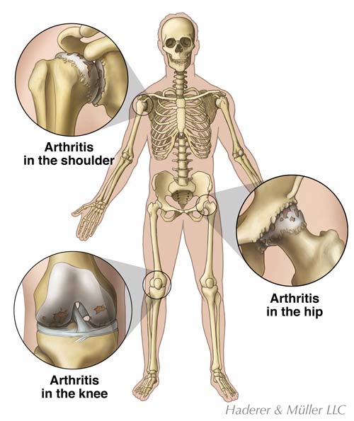
Arthritis is a common disease that causes joint pain, stiffness, immobility, and swelling.  Arthritis is actually a term for a group of over 100 diseases that affect the muscle and skeletal system, particularly the joints.  Arthritis alters the cartilage in joints.  Cartilage is a very tough, shock absorbing material that covers the ends of many of our bones.  The cartilage forms a smooth surface and allows the bones in our joints to glide easily during motion.  Arthritis can cause the cartilage to wear away.  Loss of the protective lining can cause painful bone on bone rubbing.  Arthritis can be quite painful and disabling.  While this may be tolerated with medications, therapy, other modalities, and lifestyle adjustments, there may come a time when surgical treatment is necessary.

ANATOMY

arthritis_large.jpeg

Bones are the hardest tissues in our body.  They support our body structure and meet to form joints.  Cartilage covers the ends of many of our bones and forms a smooth surface for our bones to glide on during motion.  A membrane called synovium lines the joint.  The synovium secretes a thick liquid called synovial fluid.  The synovial fluid acts as a cushion and lubricant between the joints.  It reduces friction between the bones and prevents “wear and tear.”

Ligaments are strong tissues that connect our bones together and provide stability.  Our ligaments are also lined with synovium.  The synovial fluid allows the ligaments to glide easily during movement.  Tendons are strong fibers that attach our muscles to our bones.  The tendons and muscles power the joint and enable us to move.

CAUSES

There are over 100 different types of arthritis.  Arthritis can occur for many reasons, including aging, “wear and tear,” autoimmune disease, trauma, and inflammatory disease.  Arthritis usually affects the bones and the joints; however, it can affect other parts of the body, such as muscles, ligaments, tendons, and some internal organs.  Two of the more common types of arthritis, Osteoarthritis and Rheumatoid Arthritis are discussed below.

Osteoarthritis is the most common type of arthritis, affecting some 21 million Americans alone.  It causes the cartilage covering the end of the bones to gradually wear away, resulting in painful bone on bone rubbing.  Abnormal bone growths, called spurs or osteophytes can grow in the joint.  The bone spurs add to the pain and swelling, while disrupting movement.  All of the joints may be affected by Osteoarthritis. 

Osteoarthritis is often more painful in the weight bearing joints, including the spine, hip, and knee.  It tends to develop as people grow older.  Osteoarthritis can occur in young people as the result of an injury or from overuse of a joint during sports or work.

Rheumatoid Arthritis is one of the most serious and disabling types of arthritis.  It is a long-lasting autoimmune disease that causes the synovium to become inflamed and painful.  It also causes joint swelling and deterioration.  Pain, stiffness, and swelling are usually ongoing symptoms, even during rest.

Rheumatoid Arthritis most commonly occurs in the hand and foot joints.  It can also develop in the larger joints, including the hip, knees, and elbows.  Many joints may be involved at the same time.  Further, tissues surrounding the joint may also be affected.  Rheumatoid Arthritis can affect people of all ages, but most frequently occurs in women and those over the age of 30.

SYMPTOMS

Inflammation is the main finding of arthritis.  Inflammation can cause your joints to feel painful, swollen, and stiff.  These symptoms are most likely continuous, even when you are resting.  Your joints may feel weak or unstable.  You may have difficulty moving and performing common activities, such as walking or climbing stairs.

DIAGNOSIS

Your health care provider can diagnose arthritis by conducting a physical examination.  You will be asked about your symptoms and level of pain.  Your provider will assess your muscle strength, joint motion, and joint stability.  Blood tests and other laboratory tests may identify what type of arthritis you have.  Imaging tests provide more information about the condition of your joint.

X-rays are used to see the condition of your bones and joints, and to identify areas of arthritis or bone spurs.  The tissues that surround the joint do not show up on an X-ray.  In this case, a Magnetic Resonance Imaging (MRI) scan may be requested to get a better view of the soft tissue structures, such as ligaments, tendons, and cartilage.

A bone scan may be ordered to identify the location of abnormal growths in a bone, such as bone spurs, cysts, or arthritis.  It is a sensitive test that can indicate joint degeneration in early stages that may not yet be visible on plain x-rays.  A bone scan requires that you receive a small harmless injection of a radioactive substance several hours before your test.  The substance collects in your bones in areas where the bone is breaking down or repairing itself.  These imaging tests are painless.

TREATMENT

Most cases of arthritis can be treated with non-surgical methods.  Temporary joint rest and pain relievers are sometimes all that are needed.  Over-the-counter medication or prescription medication may be used to reduce pain and swelling.  If your symptoms do not improve significantly with these medications a cortisone injection may be successful in reducing inflammation and pain.

Viscosupplementation is another injection option for arthritis, but currently is FDA approved for use only in the knee.  Studies are underway to test its usefulness in other joints.  Several products are on the market that when injected in a series into the knee can reduce pain by temporarily improving the health of the joint.

Occupational or physical therapists can help you strengthen the muscles surrounding your joint.  The resulting added joint stability can help relieve pain.  Aquatic therapy in a heated pool can be especially soothing.  In addition, the buoyancy of the water takes stress off the joints while exercising, and the resistance of the water can help strengthening efforts.  Your therapists may also apply heat to treat stiffness, and ice to decrease pain.  They may recommend splints, walkers, or canes to help relieve stress on your joints.  The therapists will instruct you on how to do your daily activities, such as housework and meal preparation, in a manner that puts less stress on your joints.

Acupuncture is a time-tested treatment for pain.  Very fine needles are strategically placed around the body to block or interrupt pain pathways.  Acupuncture should be administered by a trained professional, and can often be extremely helpful.

A variety of herbs and nutritional supplements have been shown to be helpful in treating arthritis.  Two of the more commonly known supplements are glucosamine and chondroitin.  They have been studied most in arthritis of the knee, and have shown some good results for treating arthritis in other joints. Some research has also shown that a proper diet consisting of fruits and vegetables, with a minimum of fat, can benefit arthritis.

Exercise regimes, such as yoga, Pilates, and tai chi can improve arthritis pain in many ways.  Physically, the stretching and strengthening provided by these programs has a direct positive effect for many with arthritis.  Additionally, the stress-reducing relaxation that usually occurs from these types of exercise can have a significantly positive effect on arthritis pain.

SURGERY

Because arthritis is a degenerative and progressive disease, it may get worse over time.  When non-surgical treatments no longer provide relief, surgery may be recommended.  The type of surgery that you receive will depend on your type of arthritis, its severity, and your general health.  Your doctor will discuss appropriate surgical options to help you decide what is best for you.

There are several types of surgical procedures for arthritis.  The surgeon may remove the diseased or damaged joint lining (synovium) in an operation called a synovectomy.  The bones in a joint may be realigned with a procedure called an osteotomy.  The bones in a joint may also be fused together to prevent joint motion and relieve pain.

In advanced arthritis, the damaged joint can be replaced with an artificial one.  Artificial joints are made of metal, plastic, ceramics, or a combination of the materials.  The material selected depends on the reason for the surgery, as well as which joint is being replaced.  Joint replacement can provide significant pain relief and improved mobility.

RECOVERY

Recovery from arthritis surgery is very individualized.  Your recovery time will depend on the extent of your condition, the joint that was involved, and the type of surgery that you had.  Your doctor will let you know what you may expect.

Generally, traditional open joint surgeries take several months to heal, depending on the joint.  Minimally invasive surgery and arthroscopic surgery use smaller incisions and typically heal in a shorter amount of time, from several weeks to a few months.  Your doctor may restrict your activity for a short time following your surgery.  In most cases, rehabilitation is recommended to mobilize and strengthen the joint.

PREVENTION

Some types of arthritis and arthritis symptoms may be prevented.  It is important to know what type of arthritis you have and to ask your health care provider what you can do to prevent symptoms.  For example, for some types of arthritis it is helpful to reduce your weight or stop performing repetitive joint movements.  It may be helpful to consult an occupational or physical therapist to learn how to use proper body mechanics to protect your joints during your daily activities.

Assistive devices, such as a walker, shower chair, or raised toilet seat may enable you to perform tasks while minimizing the stress on your joints.  It may also be helpful to participate in aquatic therapy in a heated pool or exercise to keep your joints strong.

FRACTURES

Bones are the hardest tissues in your body. Although bones are strong, they can split or break under too much pressure or force. A broken bone is called a fracture. Fractures can occur in a variety of ways. The most common causes of fractures are injuries, prolonged stress from overuse, and bone weakening diseases, such as Osteoporosis or tumors.

There are many types of fractures. They can range from a hairline crack to a bone that has broken into several pieces. Simple fractures may only require casting or splinting treatments. More complex fractures may need surgical intervention to align the bones for proper healing.

ANATOMY

As an adult, you have over 200 bones in your body. Your bones vary in size and shape. For instance, your arms and legs contain long bones. A series of small bones, called vertebrae, make up your spine. Very small bones form your hands and feet. Some of your bones have curves, including your ribs and skull. All of your bones line up and connect to form your skeleton. In addition to creating your body structure, your bones produce blood cells, form joints with muscles for movement, and protect your internal organs.

Your bones are live tissues. They change and grow like the other parts of your body. Most of the bones in your body are composed of the same layered materials.

The outer layer of a bone is called the periosteum. It is considered the life support system for the bone because it provides the nutrient blood for bone cells. The periosteum also produces bone-developing cells during growth or after a fracture. Underneath the periosteum is compact bone, known as the cortex. Compact bone is solid and hard. It covers the cancellous “spongy” bone. The cancellous bone looks like a sponge because it contains many pores. It can resist the stresses of weight, postural changes, and growth. In many bones, the cancellous bone contains or protects the red marrow or bone marrow. Red marrow contains developing and mature blood cells.

CAUSES

The most frequent causes of fractures are falls and motor vehicle crashes. There is a higher incidence of fractures in some sports that involve prolonged impact, high impact, balance, or high speeds. Stress fractures can result from prolonged impact or repetitive forces. For example, running or jogging can cause stress fractures in the leg, foot, ankle, or hip. High impacts can occur during contact sports, including tackles in football or punches in boxing. Skateboarding, bicycle riding, and snow skiing are sports that involve balance and speed. Fractures can occur during contact with a hard surface, for instance during a fall to the cement while skateboarding or during contact with an object, such as a tree while snow skiing.

Fractures can also be the result of physical violence. Fractures can result from a blow with a fist or kick, or from contact with a solid weapon, such as a bat.

Although the majority of fractures result from motor vehicle crashes and falls, some fractures occur because of diseases. Osteoporosis is a medical condition that causes more bone calcium to be absorbed than is replaced. Calcium is necessary for hard, healthy bones. Osteoporosis causes a reduction in bone density and brittle or fragile bones that are vulnerable to fractures. Type I Osteoporosis usually affects women between the ages of 51 and 75. Type I Osteoporosis is associated with spine and wrist fractures. Type II Osteoporosis usually affects people between the ages of 70 and 85. Type II Osteoporosis is associated with hip, pelvis, arm, and leg fractures.

Bone tumors are another disease-related cause of fractures. Most bone tumors originate elsewhere in the body and metastasize or spread to the bone. Very rarely do cancerous tumors begin in the bone. Tumors can weaken bones making them susceptible to fractures.

SYMPTOMS

In some cases, a snap or cracking sound may be heard when a bone fractures. You may feel sharp, deep, or intense pain along with numbness or tingling. Your skin may swell, bruise, or bleed.

The place where your fracture occurs may look odd, bent, or out of place. Sometimes a broken bone may come through the skin. You may not be able to move or put weight on your limb or joint, or you may do so with difficulty.

DIAGNOSIS

Your doctor can diagnose a fracture with a physical examination. Your doctor will ask you to describe your injury and your symptoms. In most cases, imaging tests are ordered to confirm the fracture.

An X-ray will be ordered to identify the type and location of your fracture. Some fractures, such as stress fractures, may not show up on an X-ray. In such cases, Computed Tomography (CT) scans or Magnetic Resonance Imaging (MRI) scans may be used to take a more detailed look at your bones. X-rays, CT scans, and MRI scans are painless procedures.

A bone scan is useful for identifying bone abnormalities from Osteoporosis or cancer. A bone scan may be used to show fractures, tumors, infection, and bone deterioration. A bone scan requires that you receive a small, harmless injection of a radioactive substance several hours before your test. The substance collects in areas where the bone is breaking down or repairing itself.

In addition to diagnosing your fracture, your doctor will classify the type of fracture that you have in order to plan treatment appropriately. Fractures are classified by a combination of general terms used to describe their features. Fractures are categorized by the characteristics of the broken bone, including the position of the fragments or broken bone and the direction of the fracture line. Common fracture characteristics and classifications are described below.

A fracture is first classified in general terms:

  • Complete Fracture: The bone is completely broken into separate pieces
  • Incomplete or Partial Fracture: A crack that does not completely break the bone into two pieces
  • Greenstick Fracture: An Incomplete Fracture with a bowed bone, it is more common in children
  • Compound or Open Fracture: The bone fragments penetrate the skin
  • Simple or Closed Fracture: The bone fragments do not penetrate the skin

Fractures are further described and classified by the position of the bone fragments:

  • Comminuted: The bones are broken into several pieces
  • Nondisplaced: The bone is broken but maintains its alignment
  • Displaced: The bone is broken and the fragments are out of position
  • Segmental: More than one fracture line leads to a "floating" segment
  • Angulated: The fragments are out of position and at an angle to each other
  • Overriding: The fragments overlap each other
  • Impacted: One piece of bone is forced into a second piece of bone.

The fracture line or crack is also described and classified. This terminology is especially used to describe fractures in the long bones of the arms and legs:

  • Linear: The fracture line is parallel with the shaft (the long part) of the bone
  • Transverse: The fracture line is at a right (90°) angle to the shaft of the bone
  • Oblique: The fracture line is at a 45° angle to the shaft of the bone
  • Spiral: The fracture line has a “corkscrewed,” “curled,” or angled pattern

SURGERY

Surgery is recommended for fractures that do not heal properly or when the bones have broken in such a way that they are unlikely to remain aligned when set with a cast. There are several options for surgery. The type of surgery that you have will depend on the location and type of your fracture. You can have general anesthesia for surgery or your doctor can numb the area with a nerve block.

Surgical options include procedures called an Open Reduction and Internal Fixation or an Open Reduction and External Fixation. Open Reduction and Internal Fixation refers to techniques that use surgical hardware to stabilize a fracture beneath the skin. Your surgeon will make an incision and place your bones in the proper position for healing. Your surgeon will secure the bones together with surgical hardware, such as rods, screws, or metal plates.

Open Reduction and External Fixation refers to techniques that use surgical hardware to stabilize a fracture from the outside of the skin. Your surgeon will make an incision and place your bones in the proper position for healing. Your surgeon will secure the bones with surgical pins that are placed through the outside of the skin. The surgical pins are attached to a metal frame on the outside of the skin.

RECOVERY

Your pain will probably cease before your fracture has completely healed. Your doctor will limit your activity while your bone is healing. Physical or occupational therapy usually follows surgery or casting. Your therapists will work with you to regain movement, strength, and flexibility that may have decreased while your bone or joint was immobile.

Recovery time from a fracture is different for everyone. It depends on the type of fracture you had and the type of treatment you received. Your doctor will let you know what to expect. Generally, fractures need about 6 weeks to heal. Some fractures can take several months to heal. Most people have good outcomes with treatment and are able to return to their regular activities.

PREVENTION

There are several things that you can do to help prevent fractures. Prevent vehicle crashes. Drive carefully. Wear a seatbelt. Make sure your vehicle is in good working order.

Prevent falls. A general physical examination can identify medical conditions that are associated with balance disorders or dizziness. Ask your doctor about a Bone Mineral Density Test to screen for Osteoporosis.

A vision exam can detect vision changes that are associated with falls. Some vision changes can be corrected with glasses. Additionally, people who wear bifocals or trifocal lenses should learn the correct way of walking with their glasses on.

It can be helpful to have an occupational therapist, a physical therapist, or a family member help you examine your home and remove obstacles that may cause you to trip. Such obstacles may include throw rugs, electrical cords, and even small pets. It may be helpful to install railings on your steps or in your shower. Wear low-heeled, sturdy shoes. They may help you maintain proper foot positioning. A cane or walker may aid your balance while you stand or walk.

If you play sports, make sure that you wear the appropriate safety equipment. Wear the safety helmets, pads, and body gear designed for your sport or activity.

Keep your bones healthy. Do not smoke. Smoking can inhibit the healing process of bones. Make sure your diet contains healthy amounts of calcium and Vitamin D. Talk to your doctor about nutrition supplements that may be appropriate for you.

SPRAINS & STRAINS

Sprains and strains are common injuries that can happen to anyone, but occur most frequently in people who participate in sports, perform repetitive activities, or are at-risk for falls.  Sprains involve the tissues that connect bones together (ligaments).  Strains involve a different group of tissues, muscles and the tendons that connect muscles to bones.  Mild sprains and strains can heal with rest and home care.  Significant sprains and strains may require rehabilitation, surgery, or both.

ANATOMY

Your skeleton is made up of bones of all sizes and shapes.  Some bones form joints that allow movement.  Muscles are strong bands of tissue that contract and relax to move bones.  Muscles are attached to bones by tendons, a fibrous tissue.  Some bones in the body are connected together by ligaments, strong tissues that provide stability and support.

CAUSES

Sprain
A sprain describes an injury to a ligament that connects two bones.  A ligament can stretch or tear if the bones in a joint move out of position from a force, such as a fall or direct contact with another person during sports.  For example, an ankle or wrist sprain can result from a fall.

Strain
A strain describes an injury to a muscle or tendon.  Muscles and tendons can be injured from overuse, overstretching, repetitive motions, sports injury, or a direct force, such as from being hit.  For example, back strain may occur in people who perform repetitive heavy lifting.

SYMPTOMS

Sprains
A sprain causes pain, bruising, and swelling.  You may hear or feel a pop when the injury occurs.  A severe sprain causes intense pain at the time of injury, followed by difficulty moving a joint.

Strains
A strain causes muscle pain, weakness, cramping, spasm, or swelling.  Moderate to severe sprains can result in muscle tearing.

DIAGNOSIS

A doctor can diagnose a sprain or strain by physical examination.  Your doctor will ask you to move your joint and test your muscle strength.  X-rays may be taken if a fracture is suspected.

TREATMENT

Immediately following your  injury, you should elevate your joint and apply cold packs to prevent swelling.  Your doctor will formulate a treatment plan based on the severity and degree of your injury.  Mild sprains and strains may benefit from physical or occupational therapy following injury.  More significant sprains and strains may require surgery or immobilization with a brace or splint for healing.

AM I AT RISK?

You may be at risk for strains and strains if:

  • Your body is deconditioned or overweight
  • You participate in sports, dance, or other challenging physical activities
  • Your job duties entail repetitive movements
  • You have experienced a strain or sprain before
  • You are at-risk for falls

COMPLICATIONS

Sprains and strains may be prevented by exercising and eating wisely to keep your body fit and healthy.  You can help prevent sprains and strains by warming up and cooling down, respectively, before and after exercising.  Be sure to wear the proper shoes and safety equipment for the sports or job duties in which you participate.  Older adults should discuss fall-risk prevention with their doctors.

TENDONITIS

Tendonitis is an inflammatory condition that can develop in a tendon. Tendons are strong fibers that connect your muscles to your bones. Tendonitis most frequently results from overuse of a joint. Symptoms include pain and tenderness. Most cases of tendonitis are relieved with rest and anti-inflammatory medications. Chronic tendonitis may require surgery if symptoms are intolerable despite nonoperative treatment.

ANATOMY

Tendons are strong fibers that connect your muscles to your bones. Tendons vary in size and shape, from the small ones in your fingers to the large ones in your legs. Your tendons and muscles move the bones in your joints.

CAUSES

Tendonitis results when the tendons are irritated in some way. Tendonitis develops because of overuse, injury, structural abnormalities, or diseases, such as arthritis and diabetes. Tendonitis commonly occurs at the shoulder, elbow, wrist, knee, and heel.

SYMPTOMS

Tendonitis causes pain and tenderness, especially near a joint. Your pain may increase with movement or activity. Pain may be present at night. The skin covering your tendon may be warm and red.

DIAGNOSIS

Your doctor can diagnose tendonitis by reviewing your medical history and conducting an examination. You should tell your doctor about circumstances that may have contributed to your condition. Your doctor will examine your muscles and tendons and will ask you to perform certain movements against resistance. There are specific examinations for each tendon.

TREATMENT

There are several different treatment options available to reduce pain and inflammation associated with tendonitis. A splint, cast, or brace may be necessary to allow your tendons to rest and heal. Your doctor may recommend using an ice pack, anti-inflammatory medications, or cortisone injections. Physical or occupational therapy can help to restore function and prevent future injury.

SURGERY

Surgery may be necessary to relieve chronic changes or inflammation around a tendon. Chronic tendonitis can cause a tendon to degenerate and tear. Surgery may be required to repair tendons that rupture as a result of chronic degeneration and inflammation. Surgical techniques vary depending on the condition and location of the tendon.

RECOVERY

Recovery from tendonitis is individualized and depends on many factors. Your doctor will let you know what to expect.

PREVENTION

You may prevent tendonitis by exercising to keep your muscles strong and flexible. Stretching properly is very important.

ArthroplastY

(Total knee or hip replacement)

Arthroplasty is surgery intended to relieve pain and to restore function to an arthritic knee or hip. The most common reason for knee or hip replacement is that other treatments (weight loss, medicines, and injections) have failed to relieve arthritis-associated knee pain. The goal of knee replacement is to relieve pain, improve quality of life, and maintain or improve knee function.

The doctors at Southeast Michigan Center for Orthopedics are hip and knee experts that perform state-of-the-art surgery and communicate closely with physical therapists for effective  post-rehabilitation. The goal is to restore pain-free hip, knee, and joint motion to allow patients to enjoy life to its fullest!

    Arthroscopy

    Arthroscopic surgery, also referred to as arthroscopy, uses an arthroscope to guide the orthopedic surgeon during a procedure. An arthroscope is a thin viewing instrument with a lens and a light that enable doctors to see inside a joint without having to open the joint up.

    Arthroscopy allows orthopedic surgeons to diagnose and repair conditions at the same time with slender surgical instruments that are inserted through the arthroscope. Arthroscopic procedures only require several very small incisions.

    Advantages of arthoscopic surgery include:

    • Small incisions
    • Less bleeding
    • Less pain
    • Decreased risk of infection
    • Faster recovery time than traditional open surgery